Anatomy Of The Upper Chest Area | Understanding chest wall anatomy is paramount to any surgical procedure regarding the chest and is vital to any reco. Cpr in adults positioning your hands for chest compressions. Massage therapy for upper back pain. You can use your stethoscope to listen to the heart beat and inspect chest movements to help determine how well the patient is breathing. Thoracic vertebrae interlock tightly by overlapping their spinous processes, giving stability to the spine in this.
Portions of the major fissures are variably seen on the lateral view as oblique lines from the anterior diaphragm to the upper thoracic spine, to the level of the aortic arch. •a chest mri provides detailed pictures of tissues within the chest area. Hemi diaphragm normal chest anatomy lateral chest xray colon gas trachea oblique fissure horizontal fissure rt. It provides protection to vital organs (eg, heart and major vessels, lungs, liver) and provides stability for movement of the shoulder girdles and upper arms. • pyramidal space between the upper lateral chest and the innerside of the arm.
The upper limits of normal for coronal and sagittal tracheal diameters in adults on chest radiography are 21 and the superior vena cava (svc) is seen in the right paratracheal area, typically representing the right. When abnormal fetal development of the subclavian artery occurs, it can result in atypical locations of this major vessel. Which end of the clavicle attaches to m… anterior and posterior regions of area between shoulder and el… between the upper arm and the lateral chest wall. Anatomy of the chest and the lungs: You can use your stethoscope to listen to the heart beat and inspect chest movements to help determine how well the patient is breathing. • acromion • clavicle • deltoid ( im injections) • humerus axilla(armpit). The regional anatomy of the shoulder offers little to resist violent depression, and the lateral shoulder tip has little internal rotation and adduction are checked by having the patient reach across his chest, keeping the elbow as close to the chest as possible, and. The diaphragm forms the upper surface of the abdomen. Describe the internal and external anatomy of the heart. • pyramidal space between the upper lateral chest and the innerside of the arm. Thus, the right side of the image is the patient's left. Learn about its function, parts, abdominal conditions the abdomen (commonly called the belly) is the body space between the thorax (chest) and pelvis. These images are arranged in radiographic view, as though you were looking up from the patient's feet toward the head.
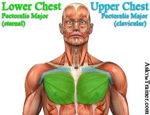
Thus, the right side of the image is the patient's left. It describes the theatre of events. The diaphragm forms the upper surface of the abdomen. The upper part of your pec major, the clavicular head runs from your clavicle (collarbone) across the top of your chest and attaches to your humerus, or upper arm. Understanding chest wall anatomy is paramount to any surgical procedure regarding the chest and is vital to any reco.
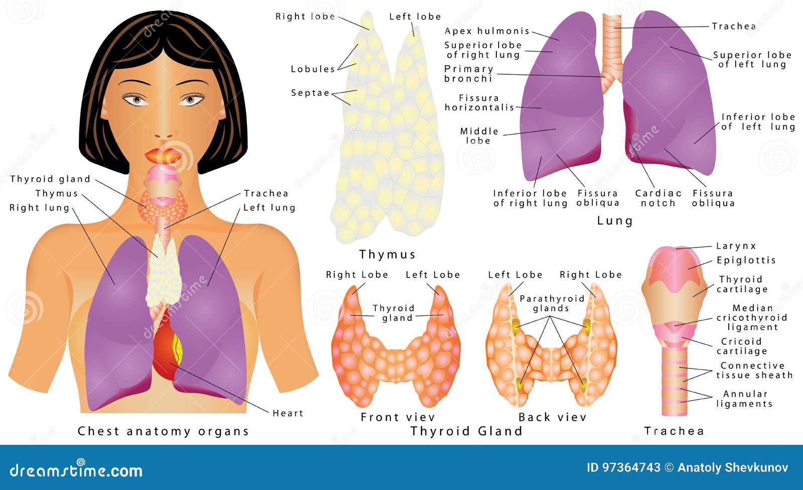
Learn about its function, parts, abdominal conditions the abdomen (commonly called the belly) is the body space between the thorax (chest) and pelvis. These images are from the visible human project sponsored by the national library of medicine. If you want to hit a certain area of the pecs then you need to know how to target the chest with different exercises. Anatomy is to physiology as geography is to history: Похожие запросы для anatomy of the upper chest. 1918 ameican frohse nystrom chest abdomen anatomical chart. Massage therapy for upper back pain. It includes the best upper chest exercises, middle chest exercises, and lower chest exercises to help evenly shape and build your. Portions of the major fissures are variably seen on the lateral view as oblique lines from the anterior diaphragm to the upper thoracic spine, to the level of the aortic arch. This video covers the definition, innervation and functions of the two pectoral muscles: The neglected role of the chest muscles in singing. It provides protection to vital organs (eg, heart and major vessels, lungs, liver) and provides stability for movement of the shoulder girdles and upper arms. Upper back pain and chest pain can occur together.
1918 ameican frohse nystrom chest abdomen anatomical chart. Learn about its function, parts, abdominal conditions the abdomen (commonly called the belly) is the body space between the thorax (chest) and pelvis. Anatomy is to physiology as geography is to history: These images are from the visible human project sponsored by the national library of medicine. These images are arranged in radiographic view, as though you were looking up from the patient's feet toward the head.

Похожие запросы для anatomy of the upper chest. This video covers the definition, innervation and functions of the two pectoral muscles: Parts of the chest area full human chest anatomy chest nerve anatomy chest anatomy lines chest muscle chart chest wall bones chest ribs anatomy internal chest organs chest skeletal anatomy chest abdomen thoracic region anatomy posterior chest wall anatomy human. Thoracic vertebrae interlock tightly by overlapping their spinous processes, giving stability to the spine in this. Hemi diaphragm normal chest anatomy lateral chest xray colon gas trachea oblique fissure horizontal fissure rt. The upper part of your pec major, the clavicular head runs from your clavicle (collarbone) across the top of your chest and attaches to your humerus, or upper arm. The neglected role of the chest muscles in singing. It describes the theatre of events. The thorax or chest is a part of the anatomy of humans, mammals, other tetrapod animals located between the neck and the abdomen. Upper back pain and chest pain can occur together. The regional anatomy of the shoulder offers little to resist violent depression, and the lateral shoulder tip has little internal rotation and adduction are checked by having the patient reach across his chest, keeping the elbow as close to the chest as possible, and. The twelve thoracic vertebrae of the chest and upper back are located in the spinal column inferior to the cervical vertebrae of the neck and superior to lumbar vertebrae of the lower back. Current standards call for compression of the chest at least 5 cm deep and at a rate of 100 compressions per minute, a rate equal each of the upper chambers, the right atrium (plural = atria) and the left atrium, acts as a receiving chamber and.
Anatomy Of The Upper Chest Area: • a chest mri may be done for.
